Introduction
The concentration of cells suspended in a sample can be determined using a specialized type of microscopy slide called a hemocytometer. It must feature a form of counting grid, the most common type being the improved Neubauer chamber, where the 3×3 mm surface is subdivided into nine 1×1mm squares. Your goal in this practical test is to determine, as accurately as possible, said concentration.
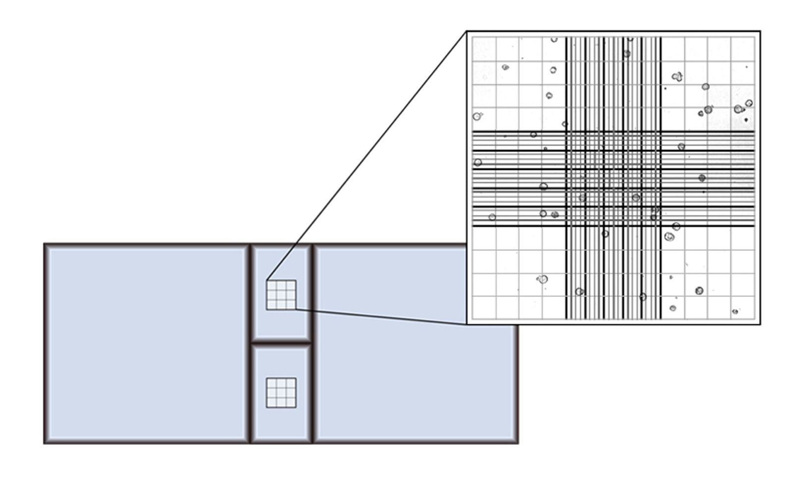
Necessary Tools and Materials
- Sample of Cells
- Microscopy stain (e.g trypan blue - used for differentiating between viable/live cells and nonviable/dead ones)
- Micropipette
- Hemocytometer
- Ethanol solution for proper cleaning of the lab equipment
- Optical microscope
Experimental method
Prepare your sample solution by adding the chosen coloring agent.
After cleaning the hemocytometer and the cover glass with ethanol and ensuring it has evaporated, pipette from the edge an exact quantity of 10 µl of your previously stained sample into one or both counting chambers. The even distribution of the suspension will be ensured by capillary action.
Place the loaded slide under the microscope and adjust the focus to optimize visibility, then count the observed cells using one of the following strategies:
- The logical count: count the cells found in the four corner squares of the grid and in the middle one. This approach will provide you with five data sets whose means are the result you need.
- The absolute count: collect data from every square on the counting grid while following a zig-zag pattern. This method is useful in high concentration samples because it provides a line of continuity that the eye can follow without getting lost.
- The quick count: take into account the concentration of cells only in the two squares diagonally opposite to each other. This method sacrifices some accuracy for speed, but it can prove useful in some cases.
TIP: Be consistent when counting the cells by establishing rules such as the cells that touch the top and right edges of a square count and the ones that come in contact with the bottom or left edges do not. This way, the risk of double counting diminishes.
Expected Results and Conclusion
The range of cell concentrations ideally determined using a hemocytometer is between \(2.5 \times 10^5\) and \(2.5 \times 10^6\) \(\text{cells}/\text{ml}^2\). As a result, you may need to dilute or concentrate your sample before counting. Keep in mind the dilution factor when calculating the concentration of the original sample.
Dilution factor = \(\frac{\text{Total volume of dilution }}{\text{Sample Volume}}\)
The total cell count can be determined using the following formula:
Total Cell Count = \(\frac{\text{Number of cells } \cdot \text{ Dilution Factor } \cdot \; 10^4}{\text{Number of squares counted on hemocytometer}} \text{cells/ml}\)
The multiplication factor of \(10^4\) in the formula above converts the count from cells per \(0.1 \; \text{mm}^3\) to cells per ml. Most hemocytometer squares have a volume of \(0.1 \; \text{mm}^3\), so the multiplication factor will be \(10^4\) in most cases.
Troubleshooting
This experiment may find its sources of inaccuracy in non-uniform cell staining, the presence of debris or procedural imprecisions such as counting errors. In order to minimize said inaccuracies, be precise when performing the dilution and pipetting steps and perform several diligent counts (typically three) with great attention and consistency (be careful what you count as a viable cell).